படிமம்:Sd4hi-unten-crop.jpg
இதைவிட அளவில் பெரிய படிமம் இல்லை.
Sd4hi-unten-crop.jpg (398 × 551 படவணுக்கள், கோப்பின் அளவு: 59 KB, MIME வகை: image/jpeg)
| இது விக்கிமீடியா பொதுக்கோப்பகத்தில் இருக்கும் ஒரு கோப்பாகும். இக்கோப்பைக் குறித்து அங்கே காணப்படும் படிம விளக்கப் பக்கத்தை இங்கே கீழே காணலாம்.
|
சுருக்கம்
| விளக்கம் |
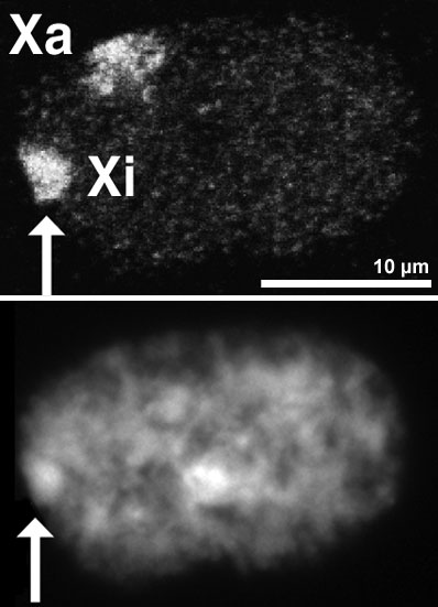
English: Nucleus of a female amniotic fluid cell. Top: Both X-chromosome territories are detected by FISH. Shown is a single optical section made with a confocal microscope. Bottom: Same nucleus stained with Dapi and recorded with a CCD camera. The Barr body is indicated by the arrow, it identifies the inactive X (Xi).
Preparation of specimen as described in: R Eils, S Dietzel, E Bertin, E Schrock, MR Speicher, T Ried, M Robert-Nicoud, C Cremer and T Cremer (1996): Three-dimensional reconstruction of painted human interphase chromosomes: active and inactive X chromosome territories have similar volumes but differ in shape and surface structure. Journal of Cell Biology, Vol 135, 1427-1440. PMID:8978813. Website.
Deutsch: Kern einer weiblichen menschlichen Zelle aus Amnionflüssigkeit. Oben: Darstellung beider X-Chromosomen durch Fluoreszenz-in-situ-Hybridisierung. Gezeigt ist ein einzelner optischer Schnitt, der mit einem konfokalen Laserscanningmikroskop erzeugt wurde. Unten: der gleiche Kern mit Dapi-Färbung, aufgenommen mit einer CCD-Kamera. Das Barr-Körperchen ist hier gut zu erkennen (Pfeil) und identifiziert das inaktive X-Chromosom (Xi).
Präparation wie in: R Eils, S Dietzel, E Bertin, E Schrock, MR Speicher, T Ried, M Robert-Nicoud, C Cremer and T Cremer (1996): Three-dimensional reconstruction of painted human interphase chromosomes: active and inactive X chromosome territories have similar volumes but differ in shape and surface structure. Journal of Cell Biology, Vol 135, 1427-1440. PMID:8978813. Website.
한국어: 여성 양수에 떠있는 세포의 핵. 위: FISH(Fluorescence in situ hybridization)법을 통해 두개의 X염색체를 볼 수 있다. 이 사진은 공초점 레이저 현미경에 의해 찍혔다. 아래 : 같은 핵을 다피 염색(DAPI)법을통해 염색하고, CCD 카메라로 찍은 사진이다. 화살표가 가리키는 것이 바소체이고, Xi로 표시된 것이 불활성화된 X염색체이다(inactive X (Xi).
사진에 관한 내용: R Eils, S Dietzel, E Bertin, E Schrock, MR Speicher, T Ried, M Robert-Nicoud, C Cremer and T Cremer (1996): Three-dimensional reconstruction of painted human interphase chromosomes: active and inactive X chromosome territories have similar volumes but differ in shape and surface structure. Journal of Cell Biology, Vol 135, 1427-1440. PMID:8978813. Website. |
| நாள் | |
| மூலம் | Steffen Dietzel, Dissertation an der Universität Heidelberg, 1996. (சொந்த முயற்சி) |
| ஆசிரியர் | User:Dietzel65, Steffen Dietzel |
| அனுமதி (இக்கோப்பை மீண்டும் பயன்படுத்துதல்) |
இந்த ஆக்கத்தின் காப்புரிமையாளரான நான் இதனைப் பின்வரும் உரிமத்தின் கீழ் வெளியிடுகின்றேன்: இந்த கோப்பு Creative Commons Attribution-Share Alike 3.0 Unported உரிமத்தின் கீழ் உள்ளது.
|
கோப்பின் வரலாறு
குறித்த நேரத்தில் இருந்த படிமத்தைப் பார்க்க அந்நேரத்தின் மீது சொடுக்கவும்.
| நாள்/நேரம் | நகம் அளவு சிறுபடம் | அளவுகள் | பயனர் | கருத்து | |
|---|---|---|---|---|---|
| தற்போதைய | 16:53, 1 அக்டோபர் 2008 | | 398 × 551 (59 KB) | Dietzel65 | {{Information |Description={{en|1=(information to be completed)}} {{de|1=Kern einer weiblichen menschlichen Zelle aus Amnionflüssigkeit. Oben: Darstellung beider X-Chromosomen durch Fluoreszenz-in-situ-Hybridisierung. gezeigt ist ein einzelner optischer |
கோப்பு பயன்பாடு
இப் படிமத்துக்கு இணைக்கப்பட்டுள்ள பக்கங்கள் எதுவும் இல்லை.
கோப்பின் முழுமையான பயன்பாடு
கீழ்கண்ட மற்ற விக்கிகள் இந்த கோப்பை பயன்படுத்துகின்றன:
- als.wikipedia.org-திட்டத்தில் இதன் பயன்பாடு
- ar.wikipedia.org-திட்டத்தில் இதன் பயன்பாடு
- bg.wikipedia.org-திட்டத்தில் இதன் பயன்பாடு
- bs.wikipedia.org-திட்டத்தில் இதன் பயன்பாடு
- ca.wikipedia.org-திட்டத்தில் இதன் பயன்பாடு
- de.wikipedia.org-திட்டத்தில் இதன் பயன்பாடு
- en.wikipedia.org-திட்டத்தில் இதன் பயன்பாடு
- et.wikipedia.org-திட்டத்தில் இதன் பயன்பாடு
- fa.wikipedia.org-திட்டத்தில் இதன் பயன்பாடு
- fi.wikipedia.org-திட்டத்தில் இதன் பயன்பாடு
- ga.wikipedia.org-திட்டத்தில் இதன் பயன்பாடு
- gl.wikipedia.org-திட்டத்தில் இதன் பயன்பாடு
- hy.wikipedia.org-திட்டத்தில் இதன் பயன்பாடு
- it.wikipedia.org-திட்டத்தில் இதன் பயன்பாடு
- ja.wikipedia.org-திட்டத்தில் இதன் பயன்பாடு
- ko.wikipedia.org-திட்டத்தில் இதன் பயன்பாடு
- mk.wikipedia.org-திட்டத்தில் இதன் பயன்பாடு
- ml.wikipedia.org-திட்டத்தில் இதன் பயன்பாடு
- ru.wikipedia.org-திட்டத்தில் இதன் பயன்பாடு
- sh.wikipedia.org-திட்டத்தில் இதன் பயன்பாடு
- sr.wikipedia.org-திட்டத்தில் இதன் பயன்பாடு
- su.wikipedia.org-திட்டத்தில் இதன் பயன்பாடு
- ta.wiktionary.org-திட்டத்தில் இதன் பயன்பாடு
- th.wikipedia.org-திட்டத்தில் இதன் பயன்பாடு
- tl.wikipedia.org-திட்டத்தில் இதன் பயன்பாடு
- uk.wikipedia.org-திட்டத்தில் இதன் பயன்பாடு
- vi.wikipedia.org-திட்டத்தில் இதன் பயன்பாடு
- www.wikidata.org-திட்டத்தில் இதன் பயன்பாடு
- zh.wikipedia.org-திட்டத்தில் இதன் பயன்பாடு